-
货号:CSB-MA000071M0m
-
规格:¥600
-
图片:
-

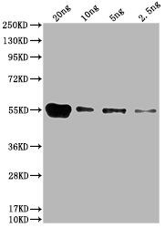
Western Blot
Positive WB detected in: 15μg hela whole cell lysate
GAPDH antibody at 1:100000, 1:200000, 1:400000, 1:800000, 1:1600000
Secondary
Goat polyclonal to mouse IgG at 1/50000 dilution
Predicted band size: 36 KDa
Observed band size: 36 KDa
Exposure time: 5min -

Western Blot
Positive WB detected in: Hela whole cell lysate at 10μg, 5μg, 2.5μg, 1.25μg, 0.625μg, 0.3125μg
All lanes: GAPDH antibody at 1:5000
Secondary
Goat polyclonal to mouse IgG at 1/50000 dilution
Predicted band size: 36 KDa
Observed band size: 36 KDa
Exposure time: 5min -

Western Blot
Positive WB detected in: Hela whole cell lysate, HepG2 whole cell lysate, Jurkat whole cell lysate, MCF-7 whole cell lysate
All lanes: GAPDH antibody at 1:2000
Secondary
Goat polyclonal to mouse IgG at 1/50000 dilution
Predicted band size: 36 KDa
Observed band size: 36 KDa
Exposure time: 30s -

Western Blot
Positive WB detected in: U87 whole cell lysate, PC3 whole cell lysate, 293 whole cell lysate, U251 whole cell lysate, A549 whole cell lysate, A375 whole cell lysate, MG-63 whole cell lysate
All lanes GAPDH antibody at 1:5000
Secondary
Goat polyclonal to mouse IgG at 1/50000 dilution
Predicted band size: 36 KDa
Observed band size: 36 KDa
Exposure time: 30s -

Western Blot
Positive WB detected in: Rat heart tissue, Rat kidney tissue, Rat skeletal muscle tissue, Rat liver tissue, Rat brain tissue tissue, Rat spleen tissue
All lanes GAPDH antibody at 1:1000
Secondary
Goat polyclonal to mouse IgG at 1/50000 dilution
Predicted band size: 36 KDa
Observed band size: 36 KDa
Exposure time: 1min -

Western Blot
Positive WB detected in: Rabbit heart tissue, Rabbit liver tissue,Rabbit spleen tissue, Rabbit lung tissue, Rabbit kidney tissue, Rabbit small intestine tissue, Rabbit skeletal muscle tissue
All lanes GAPDH antibody at 1:5000
Secondary
Goat polyclonal to mouse IgG at 1/50000 dilution
Predicted band size: 36 KDa
Observed band size: 36 KDa
Exposure time: 5min -

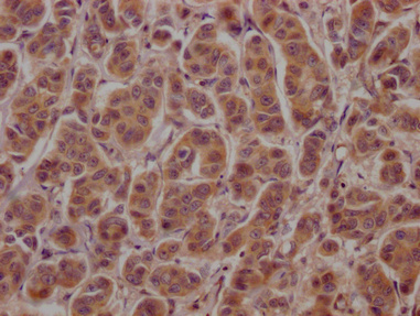
IHC image of CSB-MA000071M0m diluted at 1:100 and staining in paraffin-embedded human colon cancer performed on a Leica BondTM system. After dewaxing and hydration, antigen retrieval was mediated by high pressure in a citrate buffer (pH 6.0). Section was blocked with 10% normal goat serum 30min at RT. Then primary antibody (1% BSA) was incubated at 4°C overnight. The primary is detected by a biotinylated secondary antibody and visualized using an HRP conjugated SP system.
-

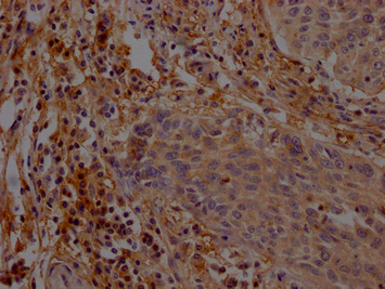
IHC image of CSB-MA000071M0m diluted at 1:100 and staining in paraffin-embedded human breast cancer performed on a Leica BondTM system. After dewaxing and hydration, antigen retrieval was mediated by high pressure in a citrate buffer (pH 6.0). Section was blocked with 10% normal goat serum 30min at RT. Then primary antibody (1% BSA) was incubated at 4°C overnight. The primary is detected by a biotinylated secondary antibody and visualized using an HRP conjugated SP system.
-

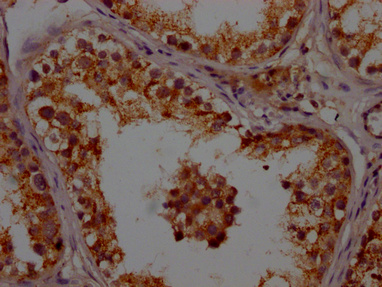
IHC image of CSB-MA000071M0m diluted at 1:100 and staining in paraffin-embedded human kidney tissue performed on a Leica BondTM system. After dewaxing and hydration, antigen retrieval was mediated by high pressure in a citrate buffer (pH 6.0). Section was blocked with 10% normal goat serum 30min at RT. Then primary antibody (1% BSA) was incubated at 4°C overnight. The primary is detected by a biotinylated secondary antibody and visualized using an HRP conjugated SP system.
-

Immunofluorescence staining of Hela cells with CSB-MA000071M0m at 1:220, counter-stained with DAPI. The cells were fixed in 4% formaldehyde, permeabilized using 0.2% Triton X-100 and blocked in 10% normal Goat Serum. The cells were then incubated with the antibody overnight at 4°C. Nuclear DNA was labeled in blue with DAPI. The secondary antibody was FITC-conjugated AffiniPure Goat Anti-Mouse IgG(H+L).
-

Immunofluorescence staining of HepG2 cells with CSB-MA000071M0m at 1:220, counter-stained with DAPI. The cells were fixed in 4% formaldehyde, permeabilized using 0.2% Triton X-100 and blocked in 10% normal Goat Serum. The cells were then incubated with the antibody overnight at 4°C. Nuclear DNA was labeled in blue with DAPI. The secondary antibody was FITC-conjugated AffiniPure Goat Anti-Mouse IgG(H+L).
-

Immunoprecipitating GAPDH in Hela whole cell lysate
Lane 1: Mouse control IgG instead of CSB-MA000071M0m in Hela whole cell lysate.
Lane 2: CSB-MA000071M0m (1μl) + Hela whole cell lysate (500μg)
Lane 3: Hela whole cell lysate (20μg)
For western blotting, the blot was detected with CSB-MA000071M0m at 1:5000, and a HRP-conjugated Protein G antibody was used as the secondary antibody at 1:2000 -

Overlay histogram showing Hela cells stained with CSB-MA000071M0m (red line). The cells were fixed with 70% Ethylalcohol (18h) and then permeabilized with 0.3% Triton X-100 for 2 min. The cells were then incubated in 10% normal goat serum to block non-specific protein-protein interactions followed by the antibody (1:200/1*106cells) for 1 h at 4°C. The secondary antibody used was FITC goat anti-mouse IgG(H+L) at 1/100 dilution for 30min at 4°C. Isotype control antibody (green line) was mouse IgG1 (1:200/1*106cells) used under the same conditions. Acquisition of >10,000 events was performed.
-

Overlay histogram showing Jurkat cells stained with CSB-MA000071M0m (red line). The cells were fixed with 70% Ethylalcohol (18h) and then permeabilized with 0.3% Triton X-100 for 2 min. The cells were then incubated in 10% normal goat serum to block non-specific protein-protein interactions followed by the antibody (1:200/1*106cells) for 1 h at 4°C. The secondary antibody used was FITC goat anti-mouse IgG(H+L) at 1/100 dilution for 30min at 4°C. Isotype control antibody (green line) was mouse IgG1 (1:200/1*106cells) used under the same conditions. Acquisition of >10,000 events was performed.
-
 1.Exosomes extracted from HEPG2 cells
2. Exosomes extracted from PC-3 cells
3. Exosomes extracted from Hela cells
4. Exosomes extracted from U87 cells
5. Hela cell Lysate -
 1.Exosomes extracted from MG63 cells
2. Exosomes extracted from Ntera-2 cells
3. MG63 cell Lysate -
 1. Exosomes extracted from Raji cells
2. Exosomes extracted from U251 cells
3. Raji cell Lysate
-
-
其他:
产品详情
-
产品描述:
The GAPDH Monoclonal Antibody is a specific antibody that targets GAPDH. The GAPDH antibody is an internal reference antibody that functions as a loading control to ensure equal protein loading and accurate quantification of protein expression levels in different samples. This antibody can detect GAPDH in human, mouse, and rabbit species.
The immunogen used to generate this GAPDH antibody is the 2-335 amino acid region of recombinant Human GAPDH protein. The GAPDH Monoclonal Antibody is raised in mouse and belongs to the IgG1 isotype. It is purified using Protein G and reaches a purity level of greater than 95%.
The GAPDH Monoclonal Antibody is available in liquid form and has been tested in various applications, including ELISA, WB, IHC, IP, and IF. These applications make the antibody a versatile tool for the detection and analysis of GAPDH in different contexts.
Moreover, the GAPDH Monoclonal Antibody has been cited in a paper by H Miao, et al. in 2022, which highlights its utility in scientific research. The use of this validated antibody in research increases the reliability of the results and ensures reproducibility.
-
产品名称:Mouse anti-Homo sapiens (Human) GAPDH Monoclonal antibody
-
Uniprot No.:
-
基因名:GAPDH
-
别名:GAPDH; G3PD; GAPD; MGC88685
-
宿主:Mouse
-
反应种属:Human, Rat, Rabbit
-
免疫原:Recombinant Human GAPDH protein (3-335AA)
-
免疫原种属:Homo sapiens (Human)
-
标记方式:Non-conjugated
-
克隆类型:Monoclonal
-
抗体亚型:IgG1
-
纯化方式:>95%, Protein G purified
-
克隆号:14C2F11
-
浓度:It differs from different batches. Please contact us to confirm it.
-
保存缓冲液:Preservative: 0.03% Proclin 300
Constituents: 50% Glycerol, 0.01M PBS, pH 7.4 -
产品提供形式:Liquid
-
应用范围:ELISA, WB, IHC, IP, IF, FC
-
推荐稀释比:
Application Recommended Dilution WB 1:5000-1:1600000 IHC 1:50-1:500 IF 1:50-1:200 IP 1µl-2µl FC 1:100-1:300 -
Protocols:
-
储存条件:Upon receipt, store at -20°C or -80°C. Avoid repeated freeze.
-
货期:Basically, we can dispatch the products out in 1-3 working days after receiving your orders. Delivery time maybe differs from different purchasing way or location, please kindly consult your local distributors for specific delivery time.
引用文献
- CXCL1 confers a survival advantage in Kaposi's sarcoma‐associated herpesvirus‐infected human endothelial cells through STAT3 phosphorylation MJ Lee,Journal of medical virology,2023
- Emerin suppresses Notch signaling by restricting the Notch intracellular domain to the nuclear membrane B Lee.et al,Biochimica et biophysica acta-molecular cell research,2017
- A fluorescence method for determination of glucose transport by intestinal BBMV of common carp Yang LP.et al,Anal Biochem,2017
产品评价

相关产品
问答及客户评论
-
Western blot review for CD146 Monoclonal AntibodyTested application: WB
Test sample: Culture cell (L-02)
Sample volume: 15μg
Primary antibody dilution ratio: 1:50,000
Review: 1:5w requires a long exposure time and is noisy. It is expected that the effect of using 1:5k should be better. Overall, the WB effect is satisfactory. It is hoped that other label antibodies with high dilution ratio can be offered for trial. -
 Western blot review for CD146 Monoclonal AntibodyTested application: WB
Test sample: Human ovarian granulosa cell carcinoma
Sample volume: 12μg, 10μg, 8μg
Primary antibody dilution ratio: 1:1200
Review: Experiment success, antibody is usable! -
 Western blot review for CD146 Monoclonal AntibodyTested application: WB
Test sample: KELLY
Sample volume: 30μg
Primary antibody dilution ratio: 1:1000
Review: GAPDH has good results in this experiment, with correct size and single band. -
Western blot review for CD146 Monoclonal AntibodyTested application: WB
Test sample: 293T Cell
Sample volume: 20μL
Primary antibody dilution ratio: 1:2000
Review: The protein is normal with clear bands, meeting the requirements of the experiment, with correct size and single bands. -
 Western blot review for CD146 Monoclonal AntibodyTested application: WB
Test sample: Human ovarian granulosa cell carcinoma
Sample volume: 10μg
Primary antibody dilution ratio: 1:1000
Review: The experiment was successful, and the antibody was successfully detected on human cells, which meets the experimental requirements and can be purchased and used in the future. -
 Western blot review for CD146 Monoclonal AntibodyTested application: WB
Test sample: Rat kidney tissue
Sample volume: 50μg
Primary antibody dilution ratio: 1:5000
Review: Good antibody, good titer. -
 Western blot review for CD146 Monoclonal AntibodyTested application: WB
Test sample: Mouse
Primary antibody dilution ratio: 1:5000
Review: Antibody specificity is very good. -
 Western blot review for CD146 Monoclonal AntibodyTested application: WB
Test sample: Human 231 cell
Primary antibody dilution ratio: 1:2000
Review: The destination band is correct and single. -
 Western blot review for CD146 Monoclonal AntibodyApplications: WB
Review: Immunoblot analyses show the presence of SGLT-1 on BBMV of common carp. Left lane: SGLT-1: BBMV. Right lane: GAPDH.
PMID: 28847591 -
 Western blot review for CD146 Monoclonal AntibodyApplications: WB
Review: HeLa cells were treated with si BAF #2 or si Control for 72 h. Cells were fractionated and subjected to western blot analyses (upper panel). Fractionation was verified by using Lamin A/C antibody (nuclear fraction) or GAPDH antibody (cytosolic fraction). The intensity of the cytosolic emerin was analyzed using ImageJ software (lower panel).
PMID: 27865926 -
 Western blot review for CD146 Monoclonal AntibodyTested application: WB
Test sample&species: Cell Lysate (Chicken)
Primary antibody dilution ratio: 1:2000
Review: Incubate at 4 °C overnight with 1: 2000 dilution ratio, the bands are clear, the specificity is good, and it can be recycled and reused with good results.

靶点详情
-
功能:Has both glyceraldehyde-3-phosphate dehydrogenase and nitrosylase activities, thereby playing a role in glycolysis and nuclear functions, respectively. Glyceraldehyde-3-phosphate dehydrogenase is a key enzyme in glycolysis that catalyzes the first step of the pathway by converting D-glyceraldehyde 3-phosphate (G3P) into 3-phospho-D-glyceroyl phosphate. Modulates the organization and assembly of the cytoskeleton. Facilitates the CHP1-dependent microtubule and membrane associations through its ability to stimulate the binding of CHP1 to microtubules. Component of the GAIT (gamma interferon-activated inhibitor of translation) complex which mediates interferon-gamma-induced transcript-selective translation inhibition in inflammation processes. Upon interferon-gamma treatment assembles into the GAIT complex which binds to stem loop-containing GAIT elements in the 3'-UTR of diverse inflammatory mRNAs (such as ceruplasmin) and suppresses their translation. Also plays a role in innate immunity by promoting TNF-induced NF-kappa-B activation and type I interferon production, via interaction with TRAF2 and TRAF3, respectively. Participates in nuclear events including transcription, RNA transport, DNA replication and apoptosis. Nuclear functions are probably due to the nitrosylase activity that mediates cysteine S-nitrosylation of nuclear target proteins such as SIRT1, HDAC2 and PRKDC.
-
基因功能参考文献:
- This suggests that RX624 might be useful as a drug against polyglutamine pathologies, and that is could be administered exogenously without affecting target cell physiology. This protective effect was validated by the similar effect of an anti-GAPDH specific antibody. PMID: 28450110
- GAPDH can interact with proteins participating in DNA repair, such as APE1, PARP1, HMGB1, and HMGB2. In this review, the functions of GAPDH associated with DNA repair are discussed in detail. PMID: 28601074
- Nitric oxide-induced GAPDH aggregation specifically induces mitochondrial dysfunction via permeability transition pore opening, leading to cell death. PMID: 28167533
- GAPDH may act as a chaperone in heme transfer to downstream areas PMID: 28315300
- NAD(+) inhibited both GAPDH aggregation and co-aggregation with GOSPEL, a hitherto undescribed effect of the coenzyme against the consequences of oxidative stress. PMID: 27282776
- Monoclonal Antibodies DSHB-hGAPDH-2G7 and DSHB-hGAPDH-4B7 Against Human Glyceraldehyde-3-Phosphate Dehydrogenase. PMID: 27556912
- the present study suggests that GAPDH plays an important role in cancer metastasis by affecting EMT through regulation of Sp1-mediated SNAIL expression. PMID: 27878251
- Knockdown of LAMP2A, a CMA-related protein, and TSG101, an mA-related protein, significantly but only partially decreased the punctate accumulation of GAPDH-HT in AD293 cells and primary cultured rat cortical neurons. PMID: 27377049
- In conclusion, the data show that two GAPDH binders could be therapeutically relevant in the treatment of injuries stemming from hard oxidative stress. PMID: 26748070
- transient silencing of GAPDH reduces intracellular ROS and facilitates increased autophagy, thereby reducing acute hypoxia and reoxygenation injury as well as the resulting apoptosis and necrosis. PMID: 26279122
- This review will summarize our current understanding of GAPDH-mediated regulation of RNA function PMID: 26564736
- analysis of PSCA level in the peripheral blood of PC patients who underwent radical prostatectomy shows it is related to a GADPH reference level (PSCA/GAPDH ratio) PMID: 26527100
- In 60 % of patients with type 2 diabetes, a reversible inhibition of GAPDH is observed. PMID: 25189828
- The results of this study led us to conclude that in cancer cells constantly exposed to conditions of oxidative stress, the protective power of Hsp70 should be abolished by specific inhibitors of Hsp70 expression. PMID: 26713364
- GAPDH and protoporphyrinogen oxidase were shown to have higher expression in faster growing cell lines and primary tumors. Pharmacologic inhibition of GAPDH or PPOX reduced the growth of colon cancer cells in vitro PMID: 25944804
- The levels of GAPDH protein were significantly up-regulated in lung squamous cell carcinoma tissues and elevated GAPDH expression is associated with the proliferation and invasion of lung and esophageal squamous cell carcinomas. PMID: 25944651
- Genetic variants in GAPDH confer susceptibility to sporadic Parkinson's disease in a Chinese Han population. PMID: 26258539
- Data revealed that GAPDH is a phosphorylation substrate for AMPK and its interaction with Sirt1 in the nucleus. The phosphorylation and the nuclear translocation of GAPDH mediate rapid Sirt1 activation and autophagy initiation under glucose deprivation. PMID: 26626483
- findings demonstrate that dissociation of the GAPDH/Siah1 pro-apoptotic complex can block high glucose-induced pericyte apoptosis, widely considered a hallmark feature of diabetic retinopathy PMID: 26438826
- Extracellular GAPDH, or its N-terminal domain, inhibited gastric cancer cell growth. GAPDH bound to E-cadherin and downregulated the mTOR-p70S6 kinase pathway. PMID: 25785838
- suggests that GAPDH aggregates accelerate Abeta amyloidogenesis, subsequently leading to mitochondrial dysfunction and neuronal cell death in the pathogenesis of AD PMID: 26359500
- The level of GAPDH-AP DNA adduct formation depends on oxidation of the protein SH-groups; disulfide bond reduction in GAPDH leads to the loss of its ability to form the adducts with AP DNA PMID: 26203648
- The activity of GAPDS was significantly positively correlated with sperm motility and negatively with the incidence of infertility. PMID: 26255202
- The N terminus of nuclear GAPDH binds with PARP-1, and this complex promotes PARP-1 overactivation both in vitro and in vivo. PMID: 25882840
- deregulated GAPDH expression promotes NF-kappaB-dependent induction of HIF-1alpha and has a key role in lymphoma vascularization and aggressiveness PMID: 25394713
- analysis of how flux through GAPDH is a limiting step in aerobic glycolysis PMID: 25009227
- astrocytic production of D-serine is modulated by glycolytic activity via interactions between GAPDH and SRR. PMID: 25870284
- Dimer and tetramer interface residues in adenine-uridine rich elements are important for GAPDH-RNA binding. PMID: 25451934
- Siah1 is a substrate of ASK1 for activation of the GAPDH-Siah1 oxidative stress signaling cascade. PMID: 25391652
- GAPDH expression is deregulated during melanoma progression. PMID: 25550585
- Oxidation of an exposed methionine instigates the aggregation of glyceraldehyde-3-phosphate dehydrogenase. PMID: 25086035
- MZF-1 binds to and positively regulates the GAPDH promoter, indicating a role for GAPDH in calcitriol-mediated signaling. PMID: 25065746
- The protein encoded by this gene contains a peptide that displays antimicrobial activity against E. coli, P. aeruginosa, and C. albicans. PMID: 22832495
- GAPDH gene over expression in resected tumor samples is an adverse prognostic factor in non small cell lung cancer. PMID: 23988223
- This review describes the structure and localization of GAPDH in cells as well as the latest discoveries on the multifunctional properties of the enzyme. PMID: 24018444
- TG2-dependent GAPDH deamidation was suggested to participate in actin cytoskeletal remodeling. PMID: 24375405
- acetylation of GAPDH (K254) is reversibly regulated by the acetyltransferase PCAF and the deacetylase HDAC5. PMID: 24362262
- GAPDH binds to active Akt, leading to Bcl-xL increase and escape from caspase-independent cell death. PMID: 23645209
- GAPDH is a moonlighting protein that functions as a glycolytic enzyme as well as a uracil DNA glycosylase. PMID: 20727968
- Results indicate that CIB1 is uniquely positioned to regulate PI3K/AKT and MEK/ERK signaling and that simultaneous disruption of these pathways synergistically induces a nuclear GAPDH-dependent cell death. PMID: 22964641
- The data presented demonstrate that up-regulation of GAPDH positively associated genes is proportional to the malignant stage of various tumors and is associated with an unfavourable prognosis. PMID: 23620736
- In a yeast two-hybrid screen of a heart cDNA library with Mst1 as bait, glyceraldehyde-3-phosphate dehydrogenase (GAPDH) was identified as an Mst1-interacting protein. PMID: 23527007
- interaction between prolyl oligopeptidase and glyceraldehyde-3-phosphate dehydrogenase is required for cytosine arabinoside-induced glyceraldehyde-3-phosphate dehydrogenase nuclear translocation and cell death PMID: 23348613
- NleB, a bacterial glycosyltransferase, targets GAPDH function to inhibit NF-kappaB activation. PMID: 23332158
- GAPDH binds to alkylated, single-stranded, double-stranded and telomeric sequences in a drug-dependent and DNA sequence/structure-dependent manner. PMID: 23409959
- GAPDH negatively regulates HIV-1 infection and provide insights into a novel function of GAPDH in the HIV-1 life cycle and a new host defense mechanism against HIV-1 infection. PMID: 23237566
- The strength, selectivity, reversibility, and redox sensitivity of heme binding to GAPDH are consistent with it performing heme sensing or heme chaperone-like functions in cells. PMID: 22957700
- The ability of C1q to sense both human and bacterial GAPDHs sheds new insights on the role of this important defense collagen molecule in modulating the immune response. PMID: 23086952
- SIRT1 functions to retain GAPDH in the cytosol, protecting the enzyme from nuclear translocation via interaction with these two proteins. PMID: 22789853
- The present mini review summarizes recent findings relating to the extraglycolytic functions of GAPDH and highlights the significant role this enzyme plays in regulating both cell survival and apoptotic death--{REVIEW} PMID: 21895736
显示更多
收起更多
-
亚细胞定位:Cytoplasm, cytosol. Nucleus. Cytoplasm, perinuclear region. Membrane. Cytoplasm, cytoskeleton.
-
蛋白家族:Glyceraldehyde-3-phosphate dehydrogenase family
-
数据库链接: